


Though the Digital Subtraction Angiography (DSA) involves the use of contrast dye, certain precautions or alternatives in contrast agents may be prescribed to patients with kidney problems to limit kidney-related harm.


Table of Contents 
Synopsis
Suffering from vascular conditions but scared of complex, invasive diagnostic procedures? Digital Subtraction Angiography can be a solution for you. A Minimally invasive diagnostic procedure not only diagnoses these conditions without large cuts and incisions but also gives precise and accurate results. Here, we will explore this modern technique along with its procedure and what conditions are diagnosed with this process.
Introduction
When diagnosing complex conditions of blood vessels in the brain, heart, and other organs, accuracy and precision are essential, which can be provided by the DSA test. The DSA is a type of angiography that can go up to the limit of precision that traditional imaging methods fail to reach. Digital subtraction angiography (DSA) can not only take clear and high-resolution images of blood vessels but also eliminate the surrounding elements like bones and other muscles, making it one of the most successful diagnostic procedures. The ability to eliminate these extra elements gains this technique its name “Digital subtraction”, meaning subtracting surrounding components digitally.
An advanced technique like DSA needs the advanced expertise of radiologists and other doctors, which is an exclusive feature of Gleneagles Hospital, a pioneer in using these modern and cutting-edge diagnostic techniques.
What is Digital Subtraction Angiography?
Digital subtraction Angiography is a fluoroscopy diagnostic technique, an imaging procedure which uses X-rays to obtain real-time images of the inside of a patient's body, which take clear images of blood vessels in a dense bundle of bone, muscles, and blood vessels. The technique is itself a great invention for diagnosis but is also used as a guide in treatment procedures for various vascular conditions. The best advantage of DSA surgery is that it enables doctors to analyse blood vessels in real-time, which allows more precision and accuracy while identifying the disease.
History of the DSA Test
To know the history of the DSA, we need to understand how and who invented the angiography itself. The development of angiography can be traced back to 1953 by Seldinger for easy access to the vascular system owing to the increasing cases of the vascular system. This invention in the 1970s proved to be a stepping stone for the invention of real-time imaging technology. Since then, it has continuously improved its clarity and accuracy of imaging by subtracting pre-contrast images from post-contrast images with digital means.
Your health matters – get expert advice today.
Why Choose Digital Subtraction Angiography
As mentioned earlier, the Digital Subtraction Angiography (DSA) is used for visualisation of various blood vessel-related issues anywhere in the body, which helps in a clearer and more accurate diagnosis of these conditions. Here are some of the most common indications of why you should choose Digital Subtraction Angiography over any other diagnostic technique:
- Endovascular Aneurysm Repair: The DSA procedure is more commonly used as a guide for different treatment procedures, such as endovascular stent grafting to treat aneurysms in emergency locations like the brain and aorta in the heart.
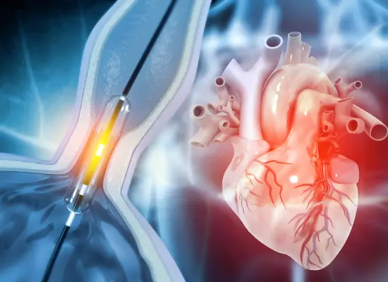
- Arterial Balloon Angioplasty: In a condition where the arteries are blocked due to various reasons, the DSA test is conducted as a guide in arterial balloon angioplasty to insert a balloon catheter or tube to widen the affected artery and restore normal functions.
- Thrombectomy: It is a condition where the blood vessels acquire blood clots. The procedure involving mechanical removal of these clots is guided by the DSA to help doctors visualise these clots easily.
- The DSA Test for Brain: The brain is the epicentre of every work we do, a complex bundle of neurons and blood vessels. These blood vessels, when malfunctioning due to various conditions like arteriovenous malformations and blood clots, can cause severe failure of not only the brain but also other organs. These conditions are visualised and diagnosed by the DSA, making it one of the most important tools in diagnosing these neurological conditions.
- Endovascular Embolisation: It is a treatment procedure used for blocking the blood vessels to treat conditions like bleeding, tumours or vascular malformations. The DSA here is used as a guide to ensure proper working of this procedure.
Preparation for DSA
Before starting the actual DSA procedure, patients will go under a thorough evaluation, which generally includes:
- Medical History Review: Your physician will review your medical histories, such as any past vascular disease, diabetes, medications, and allergies, to contrast media.
- Imaging Tests: Based on where the trouble is, your physician may have additional tests, including a CT angiogram, to get more information about your vascular disease.
- Renal Function Testing: Kidney function is evaluated to make sure your kidneys are capable of managing the contrast material employed during DSA.
- Consent and Explanation: Your doctor will describe the procedure clearly and take informed consent before starting.
The DSA Procedure
On the actual day of the conduction of the Digital Subtraction Angiography, the procedure involves the following steps:
- Acquiring the Mask Image: A series of X-ray images are first captured of the site of interest. These images record a baseline where only the contiguous anatomical structures, which are bones and other non-vascular parts, are visible.
- Injecting the Contrast Agent: After capturing the mask image, a contrast agent is administered to the blood vessels. This becomes visible on the X-ray images.
- Subtracting the Mask Image: The pre-contrast mask image is then digitally subtracted from the post-contrast images, and only the blood vessels remain visible. This subtraction removes interference from the surrounding tissues, providing a clear, detailed image of the vascular system.
- Real-Time Imaging: The subtraction images are seen in real-time so that physicians can view blood flow and any pathology, such as blockages or aneurysms. It guides further intervention, for example, stent placement or embolisation.
Recovery and Aftercare
After the procedure of DSA is done, the patient will need to rest for a few hours to a day in the hospital for observation regarding any complications. The vital signs monitored during the stay are heart rate, blood pressure, and oxygen levels. If the diagnosis involves the invasion via the groin or arm, special attention is needed for such cases to prevent complications like bleeding or hematomas.
The patients must be provided with a complete list of instructions on the activities and diet that will be needed after the DSA. The patients are also encouraged to regularly visit for follow-up and stay hydrated by drinking plenty of fluid.
Risks and Complications of DSA:
Although Digital Subtraction Angiography (DSA) is considered safe, there still are some potential risks and complications that are connected with the technique, which include:
- Bleeding & Hematoma: The most common risk connected to the DSA is bleeding at the site of catheter insertion, which results in the accumulation of blood under the skin, also known as hematoma. This bleeding leads to swelling and discomfort in the affected region.
- Pseudoaneurysm: The DSA sometimes leads to a condition known as pseudoaneurysm in which blood leaks out of a blood vessel but accumulates in the surrounding tissue. These situations need to be treated to eradicate any other complications.
- Arteriovenous Fistula: It is the connection between an artery and a vein acquired during the catheter insertion site during the DSA. This condition, when in critical areas like the brain and heart, needs surgical intervention for treatment.
- Vessel Injury: The risk of vessel damage during catheter insertion is very common, which can lead to internal bleeding.
Contrast-Induced Nephropathy (CIN): Kidney injury due to contrast dye during the DSA is quite common, particularly in patients with pre-existing kidney disease. - Thromboembolism: The development of blood clots that may be transported to another area of the body, resulting in complications such as stroke or pulmonary embolism.
- Air Embolism: The entry of air bubbles into the blood vessels that can circulate and block the blood vessels, causing hazardous obstructions.
Why Choose Gleneagles Hospitals for DSA?
When choosing a healthcare institution for Digital Subtraction Angiography, the quality of the procedure must not be compromised to ensure an effective treatment for your vascular problem. This is why Gleneagles Hospitals always provides its patients with world-class care. Here is why this premier hospital is the most trusted choice of many:
Renowned Radiologists: Gleneagles Hospital is the home of some of the most renowned and best radiologists.
Cutting-Edge Technology: The hospital is equipped with cutting-edge technology and modern infrastructure, including the latest DSA imaging systems.
Pioneers in Minimally Invasive Procedures: We pioneered using minimally invasive procedures like the DSA and many more.
High Success Rates: Gleneagles Hospital has a high success rate in performing the DSA along with the other types of angiography.

Dr Naveen Chandrashekhar
Consultant Interventional Radiologist
Qualification: MBBS, MD, FVIR (Tata Mumbai)


Dr Uma Karri
Consultant Radiologist


Dr Kushal Bhatia
Consultant
M.B.B.S., M.S. (General Surgery), M.Ch. Neurosurgery (Seth GSMC & KEMH, Mumbai) FSNVI (Fellowship in Stroke & NeuroVascular Interventions)

Frequently Asked Questions
Q1. Is the DSA procedure safe in patients with kidney problems?
Q2. What are the main indications of DSA in brain scanning?
DSA brain scanning is important in the diagnosis of vascular abnormalities in the brain, such as aneurysms, arteriovenous malformations, and other cerebrovascular diseases.
Q3. Can a brain DSA test identify stroke danger?
Yes, a DSA scan for the brain can detect cerebral vessel blockages or malformations and enable physicians to evaluate stroke risks and design preventive therapies.
Q4. How long does the DSA procedure take?
The time required for the DSA procedure varies, but it usually takes 30 minutes to an hour, depending on the complexity of the vascular condition under examination.
Q5. How does DSA compare to other types of angiography?
DSA provides better imaging definition by digitally subtracting bone and other tissues to give a better visualisation of blood vessels than standard angiography procedures.

