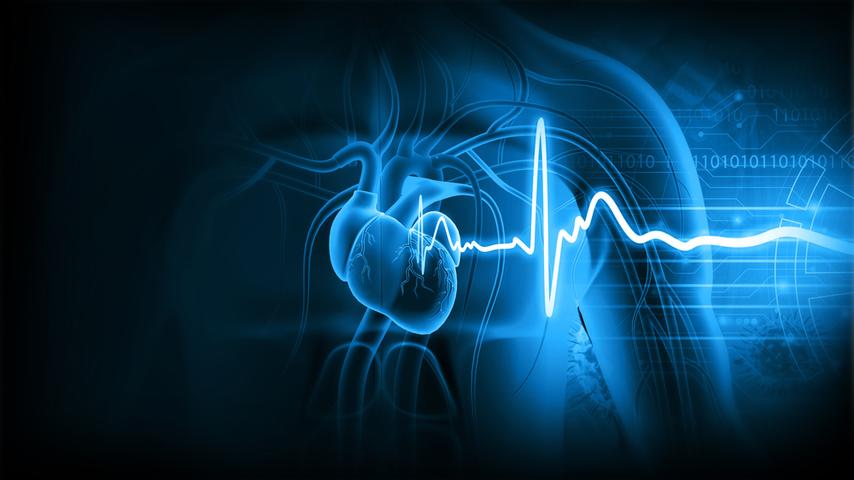
Difference Between Electrocardiogram and Echocardiography

ECG and ECHO are crucial diagnostic tests used in cardiology to evaluate the heart's function and detect abnormalities. ECG is a non-invasive procedure that records the heart's electrical activity, displaying the heart's rhythm and diagnosing irregularities like arrhythmias, conduction disorders, and myocardial ischemia.
On the other hand, echocardiography is an imaging technique that uses sound waves to create real-time images of the heart, providing detailed information about its structure, function, and blood flow. Echocardiography can assess the heart's pumping efficiency, valve condition, heart muscle thickness, and abnormalities like tumors or fluid accumulation.
ECG is quick and straightforward, while ECHO is more comprehensive and detailed, typically performed in a specialized laboratory or imaging centre. Electrocardiogram and echocardiogram both tests play crucial roles in cardiology. Due to this, the cardiologist gets a reliable way to evaluate patient's heart health. Stay tuned till the end to understand the difference between ecg and echo.
What Is An Electrocardiogram And How Does It Work?
Now that you have understood the difference between ecg vs echo. Let's delve into the functions of each. An electrocardiogram (ECG) is a diagnostic examination that captures the electrical activity of the heart, revealing important information about its rhythm, conduction pathways, and overall electrical function. An electrocardiograph is used in the test, which comprises of electrodes placed on certain parts of the patient's body.
The electrodes detect electrical signals produced by the heart with each beating, which are amplified and shown on a monitor or paper as a graph or waveform. The ECG is non-invasive and painless, taking only a few minutes to complete.
The ECG waveform is made up of many components, such as P, Q, R, S, and T waves, which correlate to different electrical events during the cardiac cycle. ECG abnormalities can suggest rhythm problems such as atrial fibrillation, conduction difficulties, myocardial ischemia, or previously occurring heart attacks. It does not provide precise information regarding structural problems or blood flow in the heart. Let's understand the process of electrocardiogram vs electrocardiograph further in the blog.
What Is An Echocardiogram and How does it work
An echocardiography is a non-invasive imaging procedure that employs ultrasonic technology to produce real-time images of the chambers, valves, and blood flow patterns of the heart. It focuses on visualising the anatomical aspects of the heart and evaluating its mechanical function, especially the size, shape, thickness, and motion of the heart muscle, as well as the integrity and performance of the heart valves.
A professional technician or cardiologist applies gel to the patient's chest and places a specialised ultrasound probe, known as a transducer, on different parts of the chest. The transducer sends out high-frequency sound waves that bounce off the structures of the heart.
These waves are translated into detailed cardiac images that are presented on a monitor. Doppler ultrasound, a technology commonly used in echocardiograms, aids in the diagnosis of diseases such as valve anomalies and intracardiac shunts.
Transthoracic echocardiography (TTE) and transesophageal echocardiography (TEE) are two forms of echocardiograms. As a diagnostic tool, echocardiograms have several advantages, including non-invasiveness, real-time pictures, and it has the capacity to identify and monitor various cardiac problems.
Differences between ECG and ECHO?
Is an ECG and echocardiogram the same? While both ECG and echocardiogram monitor the heart, they are distinct tests. An ECG identifies irregularities in the heart's electrical impulses using electrodes, while an echocardiogram uses ultrasound to examine the heart's structure for abnormalities. The difference between ecg and echo lies in what they measure and the information they provide. Electrocardiogram and electrocardiograph are terms used interchangeably to refer to the same diagnostic test. Both ecg and echo are valuable tests used in cardiology to assess the heart's health and function.
When comparing ECG and echocardiogram, they differ in terms of what they measure and the type of information they provide. Let’s understand the difference between them. The ecg vs echocardiogram tests play essential roles in cardiology, complementing each other to provide a comprehensive evaluation of the heart's health.
Electrocardiogram (ECG)
Echocardiogram (ECHO)
Records the heart's electrical activity. Focuses on the heart's electrical rhythm
Uses ultrasound to create images of the heart. Assesses the heart's structure and function
Detects rhythm abnormalities and conduction disorders
Evaluates the heart's size, shape, valves, muscle thickness, and blood flow
Non-invasive, involves placing electrodes on the body
Non-invasive, uses a specialized ultrasound probe on the chest
It can be performed by related technicians or healthcare professionals. Quick procedure, usually a few minutes
It is performed via trained cardiologist specialized technicians. It can longer time for examination
Often combined with other cardiac tests and can be performed in clinic
Specialized cardiac imaging laboratories. May be complemented by Doppler ultrasound for blood flow assessment
Initial screening tool, and preliminary assessment. Screens for electrical abnormalities.
Detailed evaluation of specific cardiac conditions. Provides detailed cardiac imaging.
It gives a snapshot of the heart's electrical activity at a specific moment
It gives real-time visualization of the heart's structures and functions
How Are ECG and ECHO Tests Done?
The ecg and echo tests are non-invasive, painless diagnostic methods used in cardiology. Moreover, the echo test vs ecg tests are conducted in clinics, hospitals, or laboratories by trained healthcare professionals. The findings contribute to the diagnosis and management of heart problems. Protocols and modifications may differ depending on the demands of the patient and the practices of the healthcare facility.
ECG Test:
The patient lies down and cleans their chest, arms, and legs. They then connect adhesive electrodes to electrocardiograph equipment. The equipment records the electrical activity in the heart and monitors the patient's breathing. The waveform is displayed on a monitor or on paper, and the data is interpreted and analyzed by a healthcare practitioner, usually a cardiologist.
ECHO Test:
The patient lies on an examination table, and a gel is applied to the chest area to facilitate the transmission of sound waves. A transducer is then placed on the chest to obtain multiple views of the heart. Ultrasound imaging captures the reflected sound waves and converts them into real-time images. Doppler ultrasound analyzes the sound waves from red blood cells to measure blood flow patterns. The results are displayed on a monitor, allowing a cardiologist to analyze the functioning of the heart.
When do you need ECGs and ECHO?
Electrocardiograms and echocardiograms are valuable diagnostic tests used in cardiology. They are employed in different scenarios to assess the heart's health and function. The need for ECGs and echocardiograms is determined by healthcare professionals based on the patient's medical history, symptoms, risk factors, and specific indications. These tests play vital roles in diagnosing and managing various cardiac conditions, enabling healthcare providers to tailor appropriate treatment plans for their patient's heart health. Let's explore when these tests are typically needed. ECGs are often performed in the following situations:
Routine Check-ups: ECGs are commonly included as part of routine check-ups, especially for individuals with risk factors such as high blood pressure, diabetes, or a family history of heart disease.
Screening for Heart Conditions: ECGs serve as initial screening tools to detect abnormal heart rhythms (arrhythmias), conduction disorders, and signs of myocardial ischemia (reduced blood flow to the heart).
Evaluating Symptoms: ECGs may be conducted when patients experience symptoms such as chest pain, shortness of breath, palpitations, or dizziness to assess the heart's electrical activity during those episodes.
Monitoring Heart Health: ECGs are used to monitor the effects of medications, evaluate the effectiveness of treatments, or assess the progress of certain cardiac conditions.
Echocardiograms are typically needed in the following scenarios:
Checking Heart Structure and Function: Echocardiograms provide detailed information about the heart's size, shape, valves, muscle thickness, and overall function. They are used to evaluate heart conditions such as heart valve disorders, congenital heart abnormalities, heart failure and so on.
Detecting and Monitoring Heart Conditions: Echocardiograms are employed to diagnose and monitor various heart conditions, including conditions affecting heart muscle function (e.g., heart failure), valve abnormalities, or congenital heart defects.
Evaluating Cardiac Symptoms: When patients present with symptoms such as shortness of breath, chest discomfort, or unexplained fatigue, echocardiograms help assess the structural and functional aspects of the heart to identify any underlying abnormalities.
Conclusion
ECGs and echocardiograms are important tests used in cardiology to check the health and functioning of the heart. ECGs keep an eye on the electrical activity and rhythm of the heart, helping doctors identify any irregular heartbeats or issues with how the electrical signals travel through the heart.
On the other hand, echocardiograms use ultrasound technology to capture real-time images of different parts of the heart. These images give doctors a wealth of information about the heart's size, shape, valves, muscle thickness, and even the patterns of blood flow.
Both tests work together to help healthcare providers diagnose heart problems, keep track of how well treatments are working, and take the best course of action for the patient's care.


